


Tests you may need, why, and what you can expect
If your signs and symptoms suggest that you have scleroderma, you’ll need to take some tests to confirm your diagnosis and narrow down the exact type of scleroderma you have. These test results show what autoantibodies you have in your blood. Autoantibodies are proteins made by your immune system that can attack your own healthy tissues by mistake.
Tests help you learn your potential disease prognosis, or long-term outlook, and your risk for specific scleroderma complications. You may only need to have these tests once at your first evaluation.
You’ll need other tests at regular clinic visits. These tests help you and your physician monitor your disease activity, or the levels of inflammation and autoantibodies in your body. Tests can also track your disease progression, or any damage to your tissues and organs that need to be addressed with treatment.
Test results help track your progress with your current treatment plan, so your rheumatologist can make changes to your therapy if needed.
Your test schedule is based on your individual clinical needs and disease type. No two people living with scleroderma are exactly alike, so you may or may not need all of these tests.
There are six categories of tests:
- ANA: Antinuclear Antibody Assay
- Nailfold Capillary Test
- Skin Tests: Modified Rodnan Skin Score
- Pulmonary (Lung) Function Tests
- Pulmonary Arterial Hypertension (PAH) Tests
- Upper GI Testing
ANA: Antinuclear Antibody Assay
Antinuclear antibody (ANA) testing is the most important blood test to screen for scleroderma and other connective tissue diseases. Your lab technician will perform a blood test, place a sample of your cells on a slide, and then examine them using a fluorescent microscope. ANA can be seen as a certain pattern of cells on the slide. The ANA autoantibodies have a fluorescent “tag” that’s easily seen under the microscope.
ANA is present in nearly all people who have scleroderma. But this test is positive in people with many other autoimmune diseases, as well as people who have no illness at all. If your ANA test is positive and your doctor also suspects you have scleroderma based on clinical signs like skin or nail changes, you will be tested for other, more specific autoantibodies too.
If you test positive for ANA and your doctor suspects you have scleroderma, they will follow up by testing you for other, more specific autoantibodies. The autoantibodies in your blood show what subset or type of scleroderma disease you have and your likely prognosis.
There are nine known scleroderma-related autoantibodies that we can detect on current commercial blood tests. Each autoantibody is seen in a particular subset of scleroderma. It is very rare to have more than one of these autoantibodies. They help your rheumatologist confirm your specific diagnosis and plan your treatment.
Three scleroderma autoantibodies are the most common. They are also seen most often in certain subsets of scleroderma, so they tell us more about your possible prognosis:
Anti-RNA polymerase III (RNA Poly III): Most often seen in people with diffuse scleroderma. People with anti-RNA polymerase III antibodies are at higher risk for rapidly progressive skin thickening and kidney problems like scleroderma renal crisis.
Anti-topoisomerase I (Topo): Also called the anti-SCL-70 antibody, more commonly seen in people with diffuse scleroderma. People with anti-topoisomerase are at high risk for developing interstitial lung disease.
Anticentromere (ACA): Most often seen in people with limited cutaneous systemic sclerosis (SSc). People with anticentromere antibodies tend to develop pulmonary arterial hypertension (PAH).
There are six other scleroderma-related autoantibodies that are far less common. They may be seen in people who also have another, overlapping connective tissue disease, like polymyositis or dermatomyositis:
- Anti-fibrillarin (U3RNP)
- Anti-Th/To
- Anti-U11/U12 RNP
- Anti-U1 RNP
- Anti-PM-Scl
- Anti-Ku
At your initial clinic appointment, you’ll also have some routine tests to check your overall health. Scleroderma can affect the function of your liver, kidney, and other organs. It’s important to have baseline testing of your organ function when you are diagnosed. You may have tests like these:
Complete blood count (CBC), which measures levels of red and white blood cells and platelets
Metabolic panel, a blood test that measures kidney and liver function
Muscle enzyme blood testing
Urinalysis, or a urine test
What to expect: Scleroderma blood and urine tests will be performed in your doctor’s office. Your nurse will tell you if you need to prepare in any way, such as skipping breakfast the day of your blood test (also called a “fasting test”). If you’re anxious about needles or having blood drawn, let your nurse or lab tech know, so they can try to minimize your discomfort or fears.
Nailfold Capillary Test
Physical changes in your nails, also known as abnormal nailfold capillaries, are one of the main symptoms in scleroderma. There are a few different machines that can test for abnormalities in the capillaries, or tiny blood vessels, inside your fingernails at the edge of your cuticles. These tests are also called nailfold capillaroscopy. They can show if your nails’ capillaries are larger than normal or bleeding slightly, all signs of vascular conditions caused by your scleroderma.
Each clinic may offer different nail tests based on the equipment they have available. Tests for nailfold capillary abnormalities include:
- Stereo microscope
- Videocapillaroscope
- Dermatoscope
What to expect: Nailfold capillaroscopy is painless. Your doctor or nurse will place your finger underneath the scope. They’ll closely examine the base of your nail for any abnormalities in the vessels.
Important preparation for your nail test: Your doctor’s office may instruct you to refrain from drinking caffeinated beverages for up to four hours beforehand. Caffeine may temporarily constrict your blood vessels, so skipping your morning coffee may help your doctor get an accurate measurement of your nail capillaries.
Also, you can’t have an accurate nailfold capillaroscopy if you’ve had any cosmetic nail treatments, such as a manicure, in the previous three weeks. Manicures or other cosmetic hand treatments can cause microscopic cracks in your nailbed that can cause false positive results on your test.
Skin Tests: Modified Rodnan Skin Score
Skin thickening, especially on the skin over your large knuckles, is one of the scleroderma symptoms that doctors look for to help confirm diagnosis. Your skin’s thickness can be measured and scored with a test called the Modified Rodnan skin score (MRSS).
This test is named after Dr. Gerald Rodnan, a scleroderma research pioneer who invented it to help measure the disease’s progressive thickening and scarring of skin, or fibrosis.
What to expect: Your doctor will closely examine several areas of your skin where you may have thickening. Usually, they will lightly pinch your skin between two fingers or a finger and a thumb to gauge its thickness on a score of zero (soft, pliable skin with no thickening) to three. This pinch will not be painful. They’ll test the skin on your hand, forearm, both upper arms, thigh, leg, both feet, face, chest, and abdomen. All the individual scores are added up for your total score.
Your Modified Rodnan Skin Score is part of a more comprehensive scleroderma test called the Composite Response Index for Systemic Sclerosis (CRISS). Your skin thickness score is added to the results of other tests that measure your lung capacity and physical function to gauge how scleroderma is affecting your body, independence, and overall quality of life.
Skin biopsy is another test to detect fibrosis, although it is not used to diagnose scleroderma. A biopsy is any test on a sample of tissue. Your doctor can do a punch biopsy, removing a small piece of your skin for examination under a microscope
and testing.
Laboratory tests on your skin samples can show signs of fibrosis, or tissue scarring. Skin changes picked up on a biopsy include overexpression of certain genes or high levels of proteins associated with scleroderma inflammation.
What to expect: Your doctor will perform a punch biopsy with a device that punches out a very small sample of skin. Typically, skin biopsy is done on a few areas of your arm, such as your upper arm, elbow and/or wrist.
First, they will clean and disinfect the areas of your skin to be biopsied. Then, they will take a regular ink pen and mark the areas where the tool will punch out each skin sample. Then, they will numb your skin with an injection of lidocaine, and a tiny amount of epinephrine to constrict the blood vessels and reduce bleeding. You will feel a little pinch and burning sensation when they give you the injections.
When they perform the punch biopsy, you should only feel some pressure, not sharp pain. Your doctor will use the tool to press down on your thick layer of skin and remove the samples to send to the lab. Your doctor or nurse will give you instructions on how to keep the biopsy areas clean, so they heal properly.
Pulmonary (Lung) Function Tests
Scleroderma may affect your lungs, causing symptoms like shortness of breath, coughing or other respiratory problems. Over time, fibrosis in your lungs can worsen, and lead to more serious lung complications of scleroderma, such as pulmonary arterial hypertension (PAH) or interstitial lung disease (ILD).
Pulmonary function testing (PFT) measures how well your lungs are working as you breathe in and out. Lung function tests may include spirometry, the six-minute walking test, and CT scans.
Spirometry is done using a machine called a spirometer that takes different measurements of your breathing and lung capacity, including:
Lung volume or tidal volume, or how much air that you take into your lungs when you breathe normally.
Diffusion capacity, which measures gas exchange in your lungs. That tells you how efficiently oxygen gas moves from the air you inhale into your lung tissue and then into your red blood cells. This is an important measurement of your lung function.
Forced vital capacity (FVC), or how much air you can forcefully breathe out after inhaling a quick, deep breath. You may also be tested for forced expiratory volume (FEV), or how much air you breathe out in the first few seconds of your FVC test.
Total lung capacity, or how much air your lungs can take in as you breathe very deeply, and vital capacity, which measures the amount of air you exhale after a deep breath.
Residual volume, or how much air is in your lungs after you breathe out as forcefully as you can manage.
Minute volume, or how much you can breathe out in the span of one minute.
Peak expiratory flow rate (PEFR), or how quickly you can breathe air out of your lungs.
Sometimes, your technician will ask you to do some gentle exercise to measure your lung volume and function during movement.
Pulmonary (meaning lung) function testing can help diagnose lung complications like PAH or ILD, even at early and more treatable stages. When you’re diagnosed with scleroderma, you’ll have a PFT to measure the baseline of your lung function. You’ll have regular PFTs to track any progression of lung disease over time.
What to expect: Pulmonary function tests are not invasive or painful. You’ll have your testing done either at your rheumatology clinic or a special pulmonary lab. Your technician will walk you through the testing. Some tests are done while you’re seated, while others are performed as you walk down a hallway in the clinic or lab.
You will breathe into a mouthpiece attached to a tube that connects to the spirometer. Some people need an adapter mouthpiece to ensure that it’s tightly fitted and doesn’t let air escape and throw off the measurements. Your lab technician will carefully instruct you on how to do the breaths for each test, including normal breathing, deep breathing, gentle breathing, or rapid breathing. They’ll give you time to catch your breath when you need to.
Six-minute walking testing measures how far you can walk during a six-minute period while your lab technician measures the oxygen saturation in your lungs, your blood pressure, and your heart rate. The six-minute walking test measures how far you can walk in meters, but not how quickly you’re able to walk. You won’t have to jog on a treadmill or do any special movements.
What to expect: While you do this test, you’ll wear a small oximeter on your finger or thumb. This small device measures oxygen in your blood. Some people with scleroderma who have Raynaud’s phenomenon, a condition where there is limited blood flow to your digits, need to wear an oximeter strapped to the forehead
Your technician will take you into a hallway or area for the test, then instruct you to walk normally back and forth between cones or marked areas on the floor. They’ll time you to stop you at six minutes. They will keep track of your heart and lung measurements during your walk.
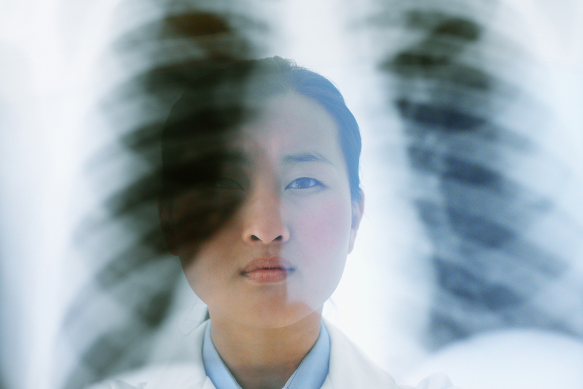
Pulmonary function testing often includes a computed tomography (CT or CAT) scan, which is a series of images taken of your lungs from different angles. CT scan creates detailed visual images of the inside of your lungs, bone structure, muscles, and tiny blood vessels in your chest.
CT scans of your lungs and chest cavity can help diagnose ILD and track any progression of disease over time. High-resolution CT scans are very detailed. They can show signs of inflammation and fibrosis in your lungs, including hazy or opaque areas, infiltrates called “ground glass,” or enlargement or swelling of the smaller airways.
What to expect: You’ll have a CT scan at the clinic, hospital, or imaging lab. The test takes anywhere from 10-30 minutes. You’ll remove your clothes and any belt, jewelry, keys, eyeglasses, or other metal objects, and put on a gown. You may be asked to drink a solution of a special dye called contrast media that highlights specific parts of your body on the CT scans. Contrast media may also be given as an injection. You’ll notice a slight warming feeling or, if you drink the solution, a mildly metallic taste in your mouth.
NOTE: Contrast media dye is iodine-based. A few people may be allergic to iodine. If you think you’re allergic to iodine, let your doctor or technician know. They will likely ask you about iodine or any other allergies during your screening for the test. If you have diabetes or any kidney problems, let your doctor or technician know before a CT scan, because your kidneys filter the iodine from the contrast dye from your blood.
For the CT scan, you will lie back and rest comfortably on a bed while your technician takes various pictures. They may ask you to hold your breath very briefly at certain points to avoid blurry images. While your CT scan is being done, you’ll hear buzzing and clicking sounds.
After your CT scan, you can go about your normal activities the rest of the day. Your technician may ask you to sit up and relax just for a few minutes just to be sure you’re feeling alright. If contrast dye was used during your scan, you’ll be given instructions on how to flush it out, including drinking plenty of fluids so you urinate.
CT scans do involve a very small amount of radiation. The amount of radiation will be kept as low as possible while ensuring that your scan results in clear images. The risk of missing signs of ILD are greater than any possible risk for this very low dose of radiation. If you’re concerned about the contrast dye or radiation involved in a CT scan, talk with your doctor.
Pulmonary Arterial Hypertension (PAH) Tests
Pulmonary arterial hypertension (PAH) is another potential complication of scleroderma. PAH is relatively rare, but it can be serious and worsen over time. It’s important to have tests that screen for and diagnose PAH very early, because it’s easier to treat at its early stages. You will also need PAH tests regularly to track your treatments’ effectiveness and any worsening of this condition.
PAH is when very small arteries inside your lungs became narrower due to inflammation. This raises the pressure of blood as it moves through these vessels—just as the term “hypertension” suggests. Your heart must work harder and harder just to pump blood through the arteries. Over time, PAH can weaken your heart.
PAH symptoms can include shortness of breath, fatigue, dizziness, swelling of your feet or legs, chest pain, and heart palpitations (sensation of your heart beating too fast).
Screening tests for PAH include:
- Physical exam
- Pulmonary function testing, particularly to measure diffusion capacity (See Pulmonary Function Test[SB1] s)
- Echocardiogram (EKG)
- Right heart catheterization
If your doctor suspects that you have PAH, you may need some or all of these tests for a diagnosis, but you won’t need them all the time.
Some people with PAH may also need other heart tests to track their heart’s function, including a stress test (done on a treadmill at a cardiologist’s office or cardiology lab) or cardiac MRI (magnetic resonance imaging, a detailed type of advanced imaging scan).
What to expect: Echocardiogram is just an ultrasound imaging scan of your heart muscle and blood flow. It is noninvasive and painless.
Your EKG can be done in your doctor’s office or clinic. Your ultrasound technician will run the ultrasound scanner attachment over your chest to take the images. They will inspect the four chambers of your heart, tissues that divide them, and valves that move blood through your heart.
Your EKG will measure the velocity or speed of blood flow through your heart and display this on a machine called an oscilloscope. This information can estimate blood pressure in your pulmonary artery. This measurement can suggest PAH, but it doesn’t confirm this diagnosis. That’s why you may need a right heart catheterization too.
Right heart catheterization is an invasive procedure, so it’s like having minor surgery. Your rheumatologist will only order a right heart catheterization if PAH is suspected and can’t be confirmed without out. This test is done at the hospital. You will be sedated. Your surgeon will insert a long, very thin tube called a catheter into your heart through one of your larger veins in your neck or leg. The catheter has a small balloon at the tip that can be inflated once it’s in the right place to measure pressure in the right atrium and ventricle of your heart, as well as the pulmonary artery.
Upper GI Testing
Scleroderma may cause gastrointestinal (GI) complications like ulcers, infections, acid reflux, heartburn and GERD, constipation, diarrhea, intestinal bleeding, or even growths and blockages. These complications can be painful and lower your quality of life. Some people with scleroderma even develop malnutrition because of damage or inflammation in their GI tract, or because they develop problems with swallowing their food, or keeping it down due to nausea and vomiting.
Upper endoscopy is a test that can diagnose GI complications of scleroderma early, so you can get treatment. An endoscope is a long, thin tube with a camera on the end that is inserted into your GI tract to show images of the interior of your esophagus, stomach, and duodenum, or the first, “upper” section of your small intestine, where these GI problems tend to occur. This test is also called esophagogastroduodenoscopy (EGD).
What to expect: EGD is a surgical procedure done at a hospital or GI procedures clinic. You’ll be sedated for the test. You won’t be awake or feel any pain or discomfort. The test takes about 20-30 minutes to complete.
Because you will be sedated for this test, you’ll need a family member or friend to drive you to and from your appointment.
Before your EGD, you’ll have an appointment with your doctor to have a physical exam and blood tests. They’ll go over any medications or supplements you take regularly, any allergies you have, if you’re pregnant or may be pregnant, and how to prepare for your test. You will be given consent forms for the test, and your doctor will explain how it works and any possible, if rare, complications.
You can’t eat or drink anything for at least eight hours before your EGD test. Once you arrive at the clinic or hospital, you’ll change into a surgical gown. An anesthesiologist will give you medicine through an IV so you go to sleep.
During the test, the endoscope is inserted into your GI tract through your mouth. Your doctor will pipe in some air to expand your stomach and intestines so the endoscope’s tiny camera can show the interior more clearly. On a screen, your doctor will look for signs of inflammation, growths, bleeding, or ulcerations. They can also take very tiny samples of tissue from your GI tract (also called biopsy) to be tested in a lab. If you have any polyps or growths of tissue, your doctor can remove them during your EGD while you sleep. Then, they’ll remove the endoscope.
Once you wake up in the recovery room, you may feel a little pressure in your stomach. You may have to flatulate to release any gas. You’ll rest for about an hour as the sedative wears off. Your nurse and doctor will tell you when it’s time to get dressed and go home. Once your friend or family member drives you home, plan to rest and relax for the remainder of the day. Test results for any biopsy done during your EGD will be ready in about a week.
Your doctor may prescribe a 24-hour GI test that you do at home on your own. This test uses a catheter inserted into your esophagus to measure acid levels and spot signs of GI complications that need to be assessed and treated, such as acid reflux.
What to expect: Your GI nurse will insert a very small, thin catheter through your nostril and into the upper part of your esophagus. You may feel a little pressure as they insert the lubricated tube. Your nurse will instruct you to swallow as they position the tube in the correct place for the test.
You go home with the catheter inserted for 24 hours. Try to do your normal activities. You can eat and drink with the catheter in place. You can eat any foods you like during meals, but only drink water between meals. Don’t fast or skip any meals.
When you lie down to sleep that night, you’ll punch a little button on a device attached to the catheter that records any symptoms you have. The next day, you’ll go back to your doctor’s office, where the nurse will remove the catheter and review the data it has collected. Your doctor can go over the test results with you and prescribe any treatments you need.
If your scleroderma causes GI symptoms like dysphagia (trouble swallowing) or pain, your doctor may suggest other tests to diagnose the cause and prescribe the right treatment. Not everyone with scleroderma will need these tests, and some tests, like colonoscopy, are standard screenings for cancer that everyone should have by the time they’re in their 40s or 50s, depending on individual risk or family history.
Esophageal dilation: During an EGD (upper endoscopy), your doctor may perform a test called esophageal dilation. They use an instrument to expand your esophagus t
Colonoscopy: If your doctor suspects that you may have intestinal bleeding, they may suggest a colonoscopy. Similar to an EGD, colonoscopy is a procedure you have while sedated. Your doctor inserts a long, thin tube with a camera on the end, but in a colonoscopy, it is inserted through your anus. The camera can show inside your colon to look for sources of bleeding. Your doctor may be able to treat the bleeding with laser therapy while you’re asleep.
Hydrogen breath test: If your doctor suspects that you have a bacterial overgrowth in your intestines, you can have a breath test to detect it. You simply drink a sugary solution, then breathe into a tube attached to an inflatable balloon. Your breath is tested for signs of gases like hydrogen or methane that are telltale byproducts of bacterial overgrowth. All you’ll need to do to prepare for this test is eat a low-carb diet for two days to cut down on the amount of fiber—which causes gas—in your system that could throw off your test results.
Gastric emptying study: This test can measure motility, or how long it takes for food to move through your GI system. If you experience bloating often, or feel very full after eating small meals, you may need a gastric emptying study to find out why. After midnight on the day of your test, don’t eat or drink anything. At the hospital, you’ll eat a breakfast that includes eggs scrambled with a radioactive solution that you won’t taste. A technician will insert an endoscope into your upper GI tract and take a series of images over four hours. You can get up and move around between each image. This test times how long it takes for your stomach to empty out the breakfast. Your doctor can interpret the results and suggest treatments if needed.
Renal biopsy: Scleroderma-related renal crisis is a rare medical emergency where blood pressure is highly elevated and kidney function declines. Signs that suggest scleroderma renal crisis include a drop in your urine production and abnormal urinalysis results. Most people with scleroderma will not need this test, but if kidney complications worsen, it is possible that you will need a renal biopsy, which would be performed surgically in a hospital.

No two scleroderma journeys are the same, but there are common experiences along the way. No matter where you, your child, or a loved one are in your journey, or the type of scleroderma, the National Scleroderma Foundation can help you find your best path.